

 It is a minimal invasive procedure that can be performed under local or general anesthesia. The procedure is acceptable for patients because it is short and together with the recovery, ends on the same day.
The field of indication of this method is very wide, covering almost all age groups of the woman after it begins to be sexually active. Indications can be the most common bleeds that are unusual or ultrasound observed changes in the cavity of the uterus and even infertility, which can be treated with this procedure with or without laparoscopy. Further it is used for contraception, for sterilization as well as for removing laid spirals or parts of them that are stale. Since the procedure is vaginal and performed locally, it is feasible to late age, or as long as there are indications.
At the same time, diagnostic and operative hysteroscopy can be performed. The diagnostic method is for observing the uterine cavity. It is usually performed under local anesthesia. It is done in the case where the exact diagnosis can not be reliably established through other physical methods such as ultrasound examination and hysterosalpingography, which have their drawbacks. Hysteroscopy provides a realistic insight wherever there is a doubt that these methods may not have discovered elements, and the image obtained from them points to a problem (local, anatomical). Operative hysteroscopy is performed under general anesthesia to treat damage inside the uterus. It can treat or remove polyps, septum in the uterus, thickening of the endometrium, or to establish that such a thing does not exist.
To perform this method, no prior preparation is required. The patient may have pre-hormonal preparation (oral contraceptives) or be made with the last days of the menstrual cycle or after its completion, when the visualization of the cavity of the uterus is almost ideal. The patient shouldn’t consume food and liquids at least 4 to 6 hours before performing the procedure and shouldn’t have anesthesiological contraindications that are very rare.
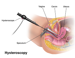
To perform the method, an optical instrument with a thickness of 3.5 mm is inserted into the uterus through the cervix. A special liquid is placed in the uterus to disperse it and give complete visualization of the entire space. The whole procedure lasts about 30 minutes, and after waking the patient is on a short observation, then she is free to go.